This essay is to be studied in relation to the two previous ones, in French, dealing with the same issue: “The Necro-Molecular Corona of Graphene” [20] and “The Name of the All-Radiant Graphene! The Molecular Spike of Graphene Oxide generates a SMOG in the human brain and in the heart of its energy system”. [21]
It is, also, to be correlated with the chapters about chitin and chitosan, in my essay, in French, “Homo chimericus: the processes of Chitinization, through insectivorous feeding, in synergy with the processes of Graphenization, will generate a new chimeric and connected human organism”. [30]
Frankly, in my youth, when I spent sleepless nights, immersed in the smoky volutes of ganja, to read the great masters of science fiction of the time – Frank Herbert, John Brunner, Philip K. Dick, etc – I would never have imagined that I would be plunged, 50 years later, into a totally dystopian universe forcing the vaccination, testing, confinement, distancing, concealment, in short, the persecution of children… legally – and with the forced encouragement of the Authorities.
All this in order to fight, allegedly, against an invisible virus… because it does not exist. What the Fake is going on?
Anybody vaccinating, testing, masking, distanciating, dissociating, or lockdowning, a child is a child abuser. Let’s call the sheriff or the local police.
Where are the Warriors, Men and Women, Mothers and Fathers, in Defense of Life?
Summary
Foreword and Declaration of Intent
Electromagnetic interference on a graphene tune
Future process improvement of biocompatible and injectable graphene oxide
Presentation of some studies with numerous Pictures of molecular necro-Coronas
Other Pictures of molecular necro-Coronas
Alleged Pictures of the invisible Sars-CoV-2
Molecular Necro-Coronas in Humans and other Animals
Molecular Necro-Coronas in Plants
Molecular Necro-Coronas in Food
Download the PDF
******************
Foreword and Declaration of Intent
My declared, and transparent, objective, for this essay, is to highlight that it is the Necro-Corona of “Graphene Oxide Molecular Spikes”, induced by the CoqueVide “vaccines”, that induces a SMOG in the human organism… and not the Spike of the invisible coronavirus… emanating from an mRNA, totally, absent from the vials of Pfizer and Moderna.
While waiting, one happy molecular day, that a Spike protein condescends to emerge/emanate, in the Biosphere – from a band of joyful amino acids, wandering about – it seems obvious, an natural, to gather, reasonably, that if such a protein does not exist much more than the Sars-CoV-2, or reciprocally, one needs to awake, then, to the sad reality of the necro-molecular Corona of graphene which is irradiating the totality of a vaccinated/injected/graphenized organism. All the more so that a lot of graphene objects have been, impeccably, Picturegraphed – by multiple researchers, around the world – while rainbowing around, under the light, in the Covid injection vials, of the Big Pharma Mafia… or, subsequently, in the organism of the Covid injected people.
I will, therefore, reiterate a small part of my recent essay [22] undermining the non-existent foundations of the pathetic arguments of a number of doctors, biologists, etc., who are dissidents of the Covidian religion. These dissidents denounce the Spikes in the Covid injections but refuse, ever, to acknowledge the nano-particular (not to say nano-technological) nature of these injections and, moreover, refuse, unfortunately, to question the vaccine dogma and the viral rantings propagated, virally, for many decades – since the official enthronement of the gigantic scams of the chemist Louis Pasteur, the Impostor.
In the USA, Ryan Cole (a dissident doctor of the Covidian religion) is convinced that if some of the injected people did not suffer from any side effects, it is because there was no mRNA, or very little, in the vials with which they were injected. And why this absence? According to Ryan Cole, because of allegedly deficient and non-compliant manufacturing processes, as well as the destruction of the mRNA induced by a non-respect of the cold chain of the vials.
It is clear that the numerous spectroscopic analyses – proving the presence of graphene – have also highlighted the total absence, or almost total absence, of mRNA in the Pfizer and Moderna CoqueVide/19 injection vials. There was no nitrogen or phosphorus.
And why would they bother putting mRNA in when they can insert graphene derivatives directly? “Vaccines” have never existed: they are one of the biggest scams – excessively lucrative – of modern allopathic medicine.
As an appetizer, I am going to ask again the questions that I have already asked – on several occasions. If, indeed, there are batches containing mRNA – because the vials have been well prepared and frozen – why not propose, then, to all the questioners that we are, Photographs, in due form, of the very famous Spike protein – that is to say, Photographs in very high definition… and reproducible. Indeed, today, thousands of studies have been published on Sars-CoV-2 without ever providing decent Photographs worthy of the name… And without providing, either, any proof of the isolation of this “virus” – by the way.
And maybe, even (one can dream), very high definition Photographs of Sars-CoV-2, the “invisible virus” – as little PharMacron called it during his historic speech declaring war… on the French People… and not on the virus.
“The narrative about the virus is the Virus”.
If this Spike protein – either natural or chimerized, in a Fauci-financed Wuhan laboratory, according to some “conspiracy theorists” – were to be really the viral and deadly bomb that is being propagated to us, why has Google never taken a very high definition picture of it?
If Google is able to propose a map of a human brain with 50,000 cells and 130 million synapses – with a virtual weight of 1.4 petabytes, i.e. the equivalent of the capacity of 700 computers [28] – it must be possible, technically speaking, to take a picture of this small, secretive virus… if it is, indeed, THE Pandemic of Pandemics. Isn’t it? Especially considering its gigantic size compared to graphene quantum dots, for example, of a few nanometers which are, nevertheless, Photographed with modern techniques.

In order to better refine this essay, I have just gone through hundreds of scientific studies (in English) dealing, only, and strictly, with the phenomenon of the “protein corona”, or “bio-corona”, or “molecular corona” – which I call “molecular necro-corona” – resulting from the fusion between nano-particles (metallic, plastic, chitosan, etc.) and the biological fluids in the human organism (blood, sperm, maternal milk, etc.)
The molecular necro-Corona is self-constructed from proteins, lipids, carbohydrates, nucleic acids.

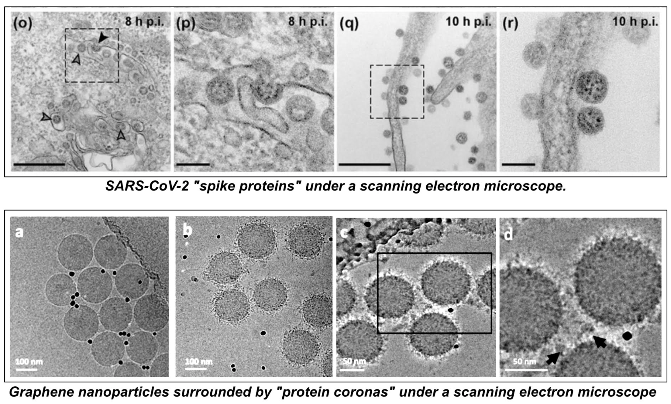
I discovered a plethora of Photographs, some of them very clear, of these “necro-molecular crowns” which represent, to a certain extent, the same objects glimpsed in a few crummy Photographs on the Web and which, supposedly, represent the invisible and enigmatic Sars-CoV-2.
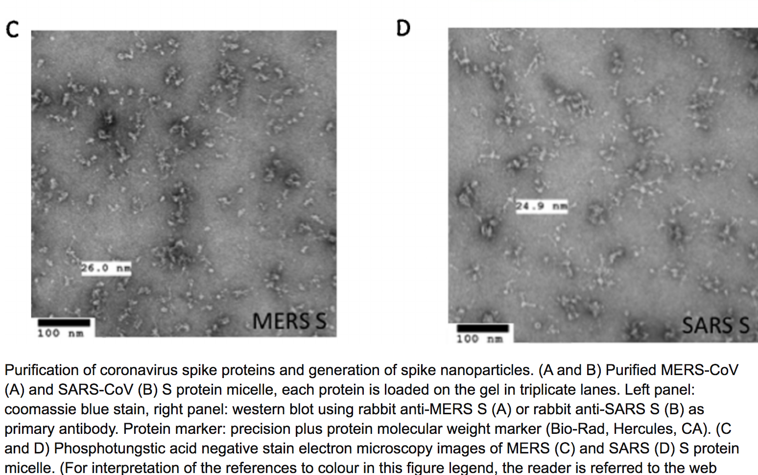
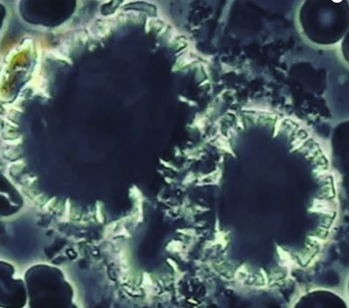
Is it necessary to specify that the size, of these alleged Sars-CoV-2 pictures, varies between 50 nanometers and 300 nanometers? Depending upon the winds of the pampa? Or is it in the nature of this coronavirus to inflate itself, like the frog of the Fable de la Fontaine, in order to confer, to itself, a semblance of importance?
The 300 nanometers picture is coming from the world expert in giant viruses, Professor Didier Raoult, who is also an expert in “Covid long”… In truth, “Long Covid” is the official denomination, issued from the Covidian religion, to cover all the symptoms generated by the nano-particles, of the graphene family, in the Covid/19 injections. Didier Raoult, a great expert in allopathy, is one of the prominent members of another line of dissidents within the Covidian religion.
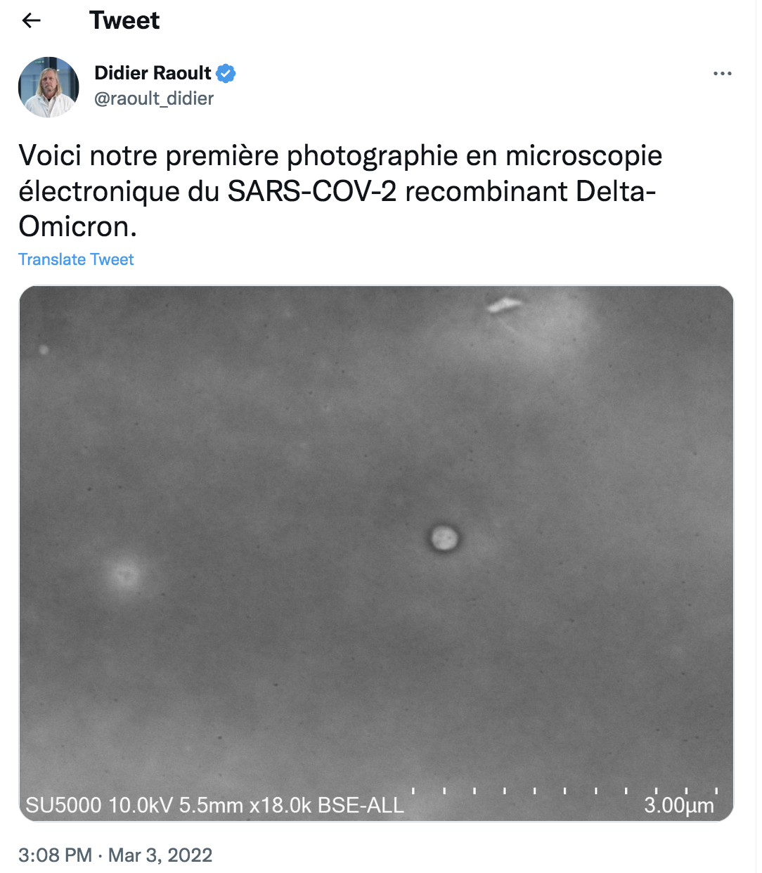
For all those who do not work with a magnifying glass, is it necessary to specify that the Delta-Omicron mutant, presented by Didier Raoult, is indeed the little round spot, encircled by black, in the middle of a huge grey rectangle? You really have to believe, blindly, in the Pasteurian theory to imagine that this little viral UFO could be a representation of the very ferocious Sars-CoV-2 which would have decimated millions of people – according to the ramblings of the corrupted technocrats of the WHO.
And to remain coherent, reasonable and rational, let’s listen to the French cardiologist Julien Devilleger (Chased by the PharMacronie and the Medical Authorities) «The photographed structure is isolated in the middle of nowhere, and does not appear to be specific at all. The image can correspond to many structures present in ANY cell or outside: endosome, lysosome, peroxisome, coated vesicle, endoplasmic reticulum, exosome… The “crown” of the “coronavirus”, is not specific, since it is found in many structures, like the exosomes for example, present in ANY cell ».
A team of researchers, from the Weizmann Institute of Science in Israel, and the University of California, calculated that each infected person would carry, at the peak of infection, approximately 1 to 100 billion virions. It is frankly bizarre that no technology has been able to capture a carrier of any of these – or a band of virions.
Moreover, when unable to photograph these little stinkers, official science has shrugged it off by saying that Sars-CoV-2 has a tolerance for size determination: from 50 nm to 140 nm. What about Didier Raoult’s giant Omicron at 300 nm? Maybe it is just a crowned nano-particle, a necro-corona, which was passing by… and which saw the light of the microscope?
No Way! This is not Sars-CoV-2! It is a necro-molecular crown on a silver nanoparticle. Science Baby!
Please click on the pictures for a better definition
Electromagnetic interference on a graphene tune
I have already dealt with this issue of “necro-molecular coronas”, in my two previous essays, but today I am going to present it again with new Photographs, new definitions and new studies, which prove, even better, the extreme danger of these chimerical abominations.
As already mentioned, I have gone through hundreds of scientific studies on this issue, but so many others are been written – for so many years – at least a thousand of them on the impact of the nano-molecular corona on human cells.
The necro-molecular corona forms around nano-materials when they are exposed to human biological fluids (blood, serum, plasma, cerebral spinal fluid, intestinal and gastric fluids, etc). The term “protein corona” was introduced, in 2007, by Tommy Cedervall and his team in a study entitled “Understanding the nano-particle/protein corona using methods to quantify exchange rates and affinities of proteins for nanoparticles.” [23]
In the case of derivatives of the graphene family, I also name this necro-molecular crown: a “Graphene Oxide Molecular Spike”.
It is this Molecular Spike of Graphene Oxide that generates a SMOG in the human brain and in the heart of its energy system. The graphenization of the human organism is totally disturbing its energetic system in order to introduce an electromagnetic jamming – a neuronal jamming… When it will not be, directly, an emission of injunctions in the heart of the human brain… in the course of the graphenizing injections: the first one, the second one, the third one, the fourth one…
I invite the readers, who do not know what the energetic body corresponds to, to consult the foundations of Traditional Chinese Medicine, Traditional Tibetan Medicine, Traditional Ayurvedic Medicine…
This electromagnetic jamming has, as a function, quite simply, to operate a Great Reset, a Great Reinitialization, in the heart of the human brain, in the heart of its nervous system and in the heart of the second brain, the heart – in short, in the heart of its whole energetic system.
This electromagnetic interference generates a huge existential mess. Question: Who is in charge, neuronally, when the brain has been graphenized?
Graphene inherently scrambles what has been given to every human animal since the dawn of time; it scrambles, thus, the fundamental source-codes – both information and genetic.
The Molecular Spike of Graphene Oxide is a vector of contamination, of parasitization, of vampirization, towards the metamorphosis – the Great Reinitialization – of the organic body into an organo-metallic body… knowing that graphene is a half-metallic and half-mineral compound. Graphene is, therefore, a chimera in many respects.
Moreover, this chimerizing SMOG often manifests itself, in terminal mode, by the constitution of very long and very thick blood clots… which, according to some studies, contain almost no blood.
The Molecular Spike of Graphene Oxide is a vector of contamination, of parasitization, of vampirization, towards the metamorphosis – the Great Reinitialization – of the organic body into an organo-metallic body… knowing that graphene is a half-metallic and half-mineral compound. Graphene is, therefore, a chimera in many respects.
This is, truly, the Great Reset, the Great Reinitialization, orchestrated by the demented eugenicist, Klaus Schwab – and all his accomplices, Bill Gates, Anthony Fauci, Tedros Adhanom Ghebreyesus, etc., etc., ad nauseam. The other, the Great Sociological Reset, is subsequent to and, in fact, dependent on the first graphene-scented Reset.
Our brains are the prime target of Graphene – and of those who want to inject it into you. And who are they? Who are they? WHO? If you can’t see it, it’s possible that your brain is already being vampirized by graphene.
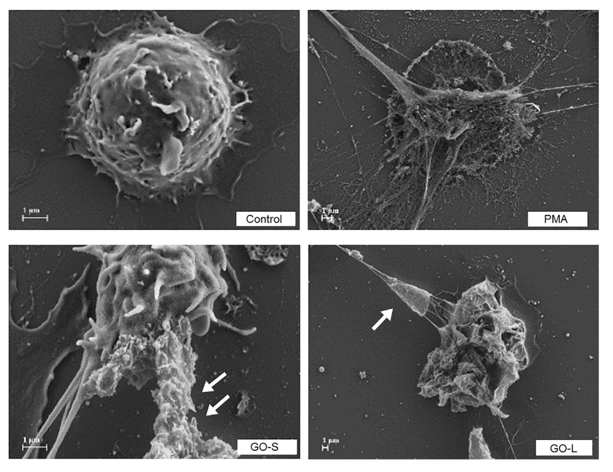
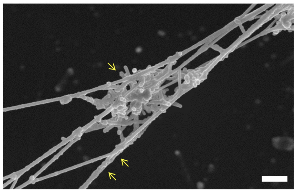
SEM micrographs of control neutrophils or neutrophils exposed to PMA (25 nM), large graphene oxide sheet (12.5 mg/mL) or small graphene oxide sheet (12.5 mg/mL). The single arrow (B) indicates a large graphene oxide sheet trapped in a network of chromatin fibers (i.e. NETs). The pair of arrows (B) indicates chromatin fibers (NETs) covered with small graphene oxide sheets. Scale bar: 1 micron.
Future process improvement of biocompatible and injectable graphene oxide
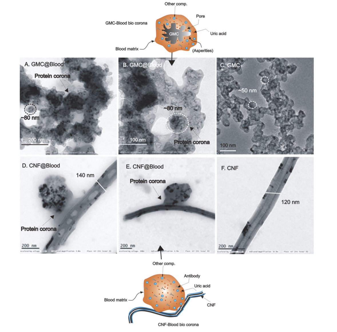
I will, first of all, represent the introduction of a study, from 2018, which announces, ostensibly and clearly, the presence of graphene in injections. It is “Graphene oxide touches blood: in vivo interactions of bio-coronated 2D materials” [29] which focuses on the bio-molecular crown of graphene and its potential for toxicity in the human body. The authors describe the journey of graphene oxide following its injection into the bloodstream: from initial interactions with plasma proteins to the formation of the bio-molecular corona and its bio-distribution.
As a reminder to all Decoders and other Fact- Checkers in the service of the Covidian Sect. This study on the potential toxicity, of the graphene oxide necro-molecular corona, was published back in October 2018. Its intention is clearly worded – at least for funding purposes.
This study is all about the development of pharmaceutical systems, based on bio-compatible graphene, injected intravenously. It is all about the creation of “bio-compatible” graphene-based vaccines. Thus, what will the ImMonde’s Decoders or the AFP’s ignorant defakers babble? Will they change the meaning of the term “biocompatible”… because, considering the intended genocide, it would be, rather, necro-compatibility!
«The in vivo fate of nanomaterials is generally influenced by several factors, including the route of administration, nanomaterial chemistry and physiological environment. Numerous physicochemical characteristics including lateral size, shape, dose, exposure time, number of layers, chemical composition, surface charge, stability, purity, and surface functionality can influence the fate after injection, when materials are exposed to the rich milieu of blood proteins. The proteins in the bloodstream cause an immediate and dramatic change in the biological ‘‘identity’’ of nanomaterials. The result is the development of a new interface, consisting of a dynamic shell of blood macromolecules. This layer, given the protein enrichment, is usually referred to as the protein corona or the biomolecular corona. The biomolecular corona determines the interactions with cells, uptake and clearance and therefore affects the biodistribution and delivery to the intended target sites.
The biomolecular corona of Graphene Oxide is still poorly explored and few works have considered the influence of this layer on in vitro and in vivo effects. Here, we will first discuss the surface features of Graphene Oxide and their links to amino acids and blood protein binding. Then, we will discuss the Graphene Oxide/biomolecular corona composition and how the biomolecular corona can influence interactions with blood cells. Finally, we will highlight biodistribution and biosafety concerns as well as future challenges related to the development of intravenously injected Graphene Oxide-based pharmaceutical systems. The comprehension of these aspects is involved in and will improve the future design of injectable biocompatible Graphene Oxide. Emphasis from Xochi. [29]
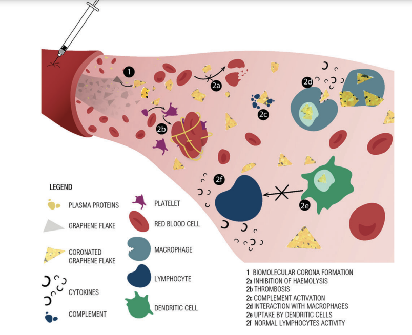
Picture 40. Above. Main results of GO interaction with blood components are summarized in this illustration of the injection of GO flakes in the bloodstream. The formation of the BC (1) prevents the hemolysis of red blood cells (2a). Thrombosis (2b) and interaction with complement proteins (2c) are ascribed to GO. In (2d) some of the possible fates after macrophage encounters are shown: extracellular blocking or intracellular uptake. The release of cytokines occurs when macrophages uptake GO. Aggregates of GO in macrophage cytoplasm induce the production of pro-inflammatory cytokines. Dendritic cells fail to present antigens to lymphocytes when they uptake GO (2e). Lymphocyte activity is not inhibited, and BC protects lymphocytes from apoptosis (2f).
This study was commented last year by the Spanish scientist Mik Andersen: “Graphene oxide in contact with blood”. [32]
The authors seem to have noted the dangers of GO, in fact they indicate that «Small GO flakes (few hundreds of nm) seem to be more destructive». Nevertheless, they argue that protein coating with BC, among other procedures, can reduce the risk of “red blood cell hemolysis,” i.e., the disintegration of red blood cells. Strategies for coating and protecting the GO so that it is tolerated by the human body are discussed in turn. With regard to this section, the risk of thromboembolism is also implicitly recognized with rGO (Reduced Graphene Oxide) and GO. In fact, it is indicated that «When administered in vivo (250 mg kg1 body weight), 48% of lung vessels were partially occluded after 15 minutes.», then, it is clarified that «This in vivo impact on the coagulation cascade can be caused by an aggregation of the nano-material after injection.».
The article confirms that «understanding the interaction of GO with immune cells is crucial for the development of biomedical technologies». This indicates the interest in applying graphene technology despite the natural rejection by the human body and the problems it generates – see references to scientific studies on graphene toxicity.
Regarding the immune response of the human body, the following statement can be quoted: «A recent study demonstrated that GO induced pro-inflammatory cytokine expression in a size-dependent fashion, with smaller sized GO (o1 mm) being more effective than larger ones (1–10 mm).». Cytokines were one of the most contrasting symptoms during the COVID-19 pandemic, which allows one to infer a causal relationship with what is reported in the article.
It is indicated that «Within 48 hours after i.v. injection, GO is cleared from the bloodstream and distributed throughout various organs with preferred accumulation in the lungs, liver, and spleen. The lack of pathological changes was reported after 14 days of treatment at a low dose (1 mg kg1 ), but at a higher dose (10 mg kg1 ), granulomatous lesions, pulmonary edema, inflammatory cell infiltration, and fibrosis throughout the lung were observed.».
This study also alludes to degradation problems that can lead to accumulation problems in cells. Indeed, it is about the « Finally, the degradation of injected GO is an important biosafety concern. Long-term interaction (14 days) of GO with plasma causes reduction and biodegradation with hole formation caused by the action of hydroxyl radicals. Once internalized by the immune cells, biodegradable particles are digested and cleared from the body, while non-biodegradable particles accumulate in cells for extended periods».The author does not allude to the negative effects that such a build-up may cause. The authors acknowledge that graphene is still far from being ready for safe clinical treatment in patients, as they state: «Despite the great scientific advances, future studies for in vivo application should focus on some weaknesses in graphene research. First, graphene materials should be designed to have more than just a stable small size for rapid excretion, and degradable composition to limit toxicity».
Presentation of some studies with numerous pictures of molecular necro-Coronas
I will now present various texts, as well as various studies, that present, in different ways, some aspects of the molecular crowns that develop during the fusion between nano-particles and the cells of the body.
Please click on the pictures for a better definition
Picture 33. Micrograph taken under Phase Contrast Microscopy reveals the live blood 24 hours after the mRNA Vaccine now containing crystallized red blood cells, biological transformations of red and white blood cells, large symplasts of reduced graphene oxide or graphene hydroxide crystals center and Orotic acid crystals in the upper right hand corner of the micrograph. Dr. Robert O. Young, Hikari Omni Publishing, September, 2021. [31]
“New Quality-Control Investigations on Vaccines: Micro- and Nanocontamination”. Antonietta M Gatti et Stefano Montanari. [1]
«The investigations revealed that some particles are embedded in a biological substrate, probably proteins, endo-toxins and residues of bacteria. As soon as a particle comes in contact with proteic fluids, a nano-bio-interaction occurs and a “protein corona” is formed. The nano-bio-interaction generates a bigger-sized compound that is not biodegradable and can induce adverse effects, since it is not recognized as self by the body.»
“Multicolor Super-Resolution Microscopy of Protein Corona on Single Nanoparticles”. [11]
According to their introduction. «The protein corona, or biomolecular corona, is a coat assembled on the surface of nanoparticles once they are administered into a biological medium containing biomolecules such as enzymes, proteins, or peptides. Upon being coated by protein corona, the nanoparticle’s biological fate will be largely determined by the protein coat rather than the underlying functionalized nanoparticle, giving a new identity that governs the cellular uptake process of the nanoparticle. Protein corona also impacts the performance of nanoparticles when used as label-free biosensors because the accessibility of surface functional groups is modulated. It is now known that the formation of protein corona is highly dynamic and dependent on a wide range of nanoparticle properties such as size, shape, and surface chemistry. Although significant progress in ensemble characterization of protein corona has been made, a deep and comprehensive understanding of protein−nanoparticle interaction at the molecular scale is vital for the development and rational design of particle-based nanomedicine and biosensors ». [11]
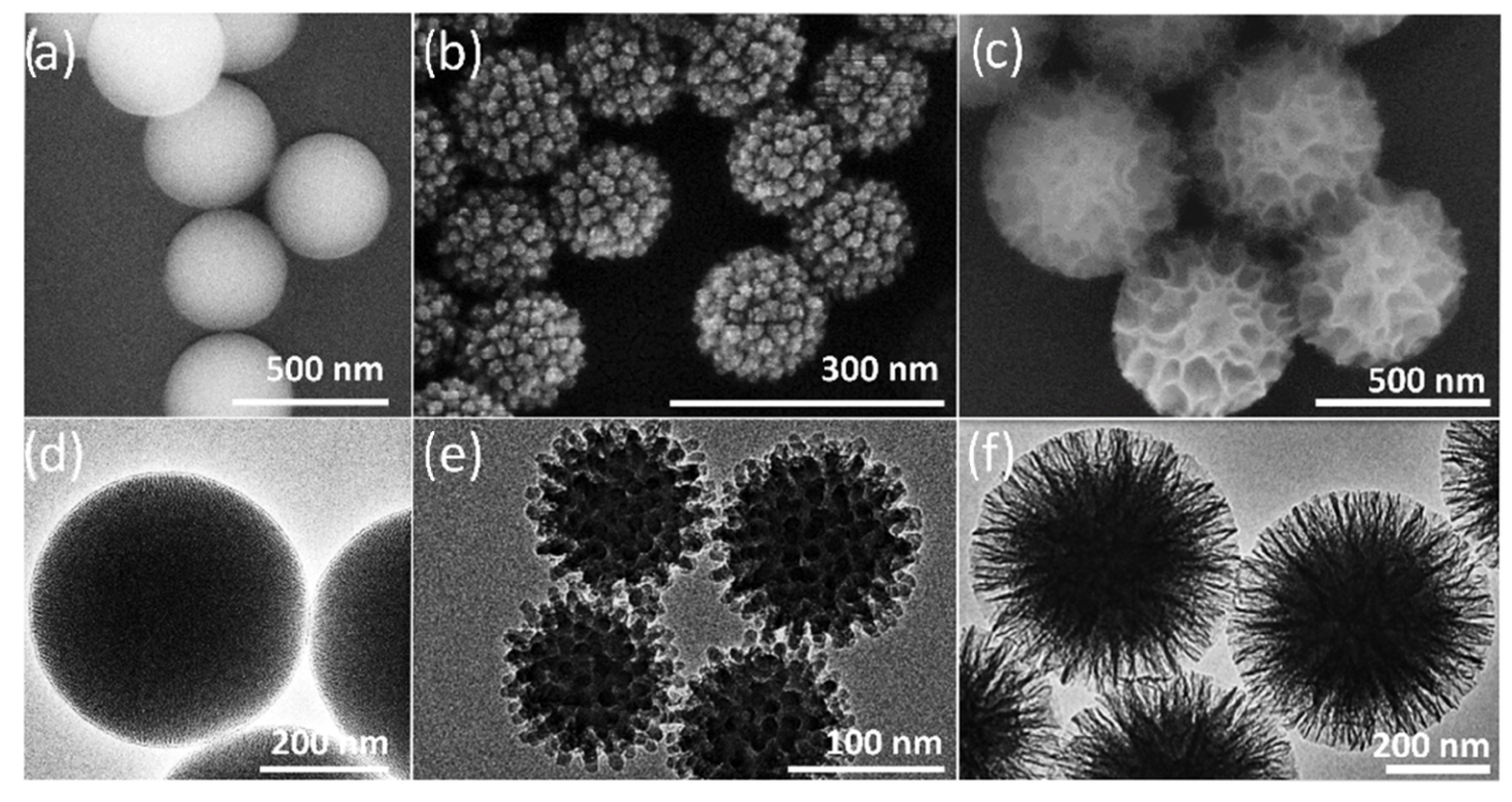
Picture 06. Above. Morphological overview of silica nanoparticles for protein corona formation, displaying (a−c) SEM and (d−f) TEM images of MSN, VSN, and WSN nanoparticles, respectively.
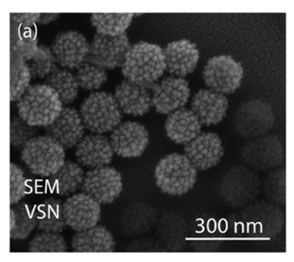
Picture 07. Above. SEM images of (a) virus-like silica nanoparticles.
“Synthesis of Patient-Specific Nanomaterials”. [33]
This study investigates the various protein crowns, in the same individual, generated by the contact of nanoparticles with various fluids: serum, breast milk, saliva and tears.
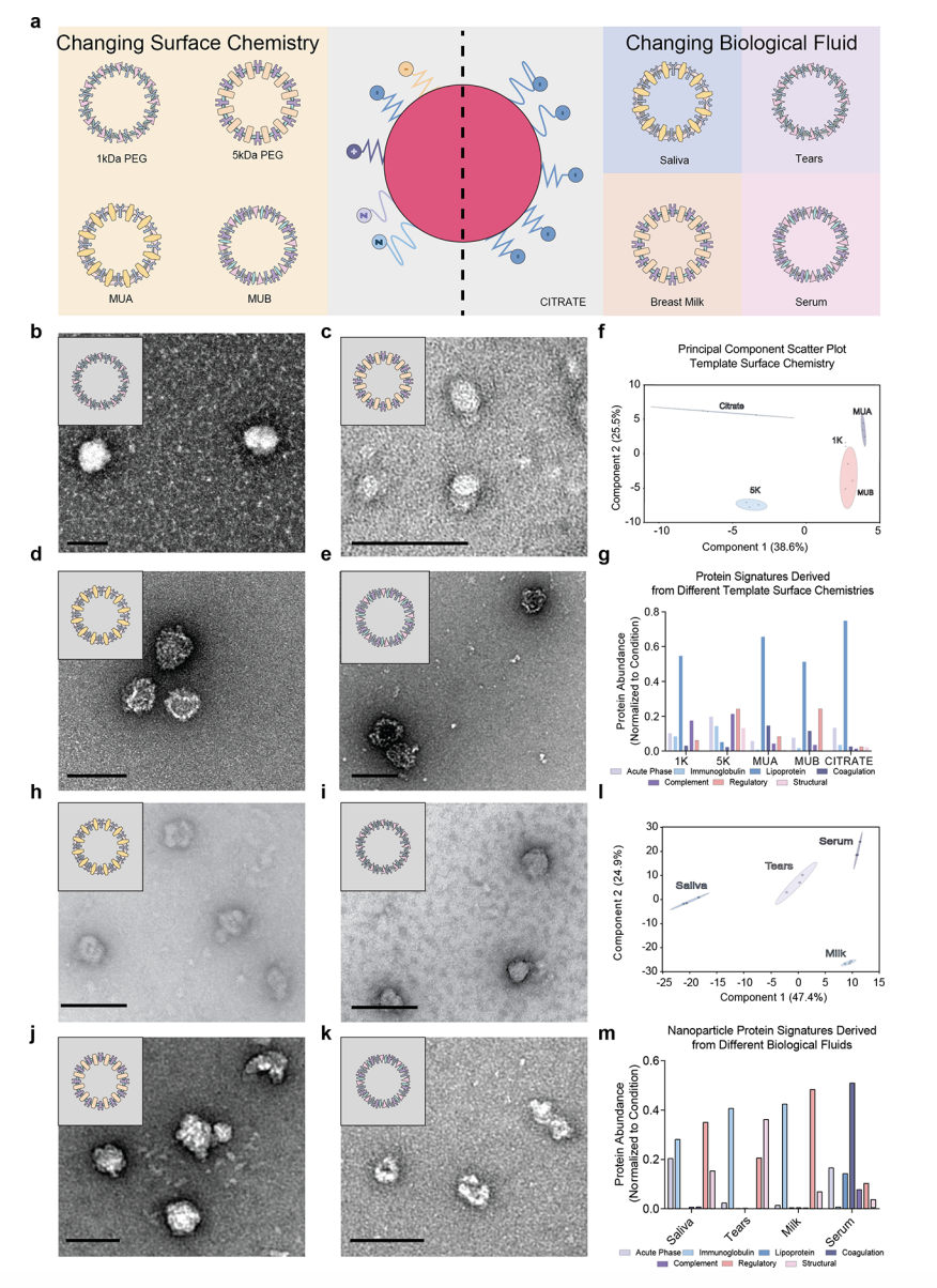
Picture 42. Scale Bar, 100 nm. Above.
Engineering protein nanoparticles by changing template surface chemistry and source biological fluid. (a) Altering template surface chemistry to anionic, neutral, or cationic changes the distinct protein compositions found in PNPs. Different template surface chemistries such as (b) mercaptoundecanoic acid (MUA), (c) mercaptoundecyltrimethylammonium bromide (MUB), (d) 1 kDa polyethylene glycol (PEG), and (e) 5 kDa PEG produced reproducible PNP. (f) Principal component analysis (PCA) derived from mass spectrometry data with biological replicates (n = 3), shows the variability and statistically unique compositions derived from the different template surface chemistries. Colored circles represent the 95% confidence region for each replicate (n = 3). The greater the overlap between regions, the more similar they are. (g) The statistically significant proteins that were discovered using the multivariable analysis of variance were classified according to their gene ontology function using the Universal Protein Database. Similarly, (h) serum, (i) breast milk, (j) saliva and (k) tears were isolated from patients and used to make PNP. Each PNP was significantly different from one another according to (l) PCA and (m) multivariable analysis of variance. Scale bar, 100 nm.
“The nanoparticle biomolecule corona: lessons learned – challenge accepted?” [7]
«Hence, albeit we are faced with more than a thousand reports on the impact of the NP corona on human cells, it is more than surprising that no studies on the relevance of the NP corona on the microbiome, as well as on microbiome–host interactions, have been reported so far».
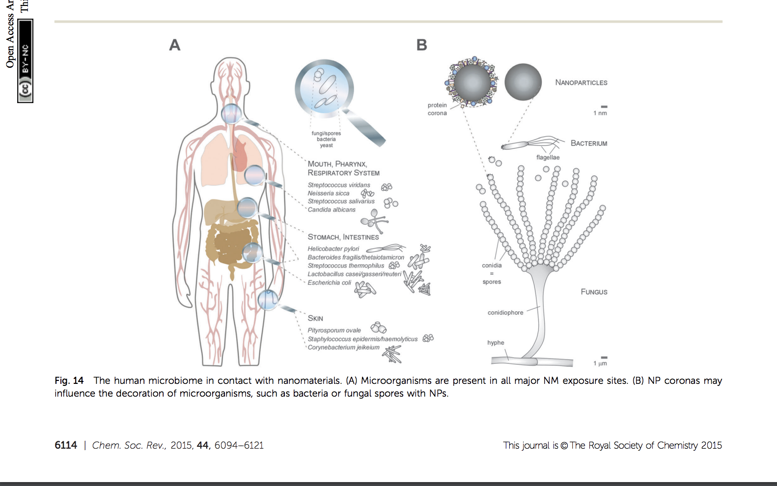
Picture 32. Above. The human microbiome in contact with nanomaterials. (A) Microorganisms are present in all major NM exposure sites. (B) NP coronas may influence the decoration of microorganisms, such as bacteria or fungal spores with NPs.
“The impact of protein corona on the biological behavior of targeting nanomedicines”. [35]
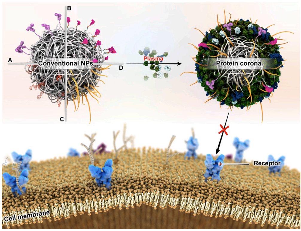

Picture 02. Above. a Original NPs without corona; b NPs with corona; c, d higher magnification of the NPs showing the distribution of biomolecules and their association with the surface of the NPs. Nonspecific proteins accumulate between the NPs as large clusters (black arrows). The black dots in the images are 10 nm gold fiducial markers.
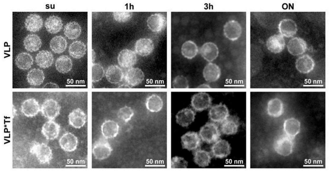
Picture 45. Above. Representative TEM images of VLP, and VLP*Tf visualized by negative staining. Images of samples without serum addition (SU), after incubation in 55% human serum for 1 h, 3 h, or overnight (ON). Adapted with permission from ref (Zackova Suchanova et al., 2020). Copyright 2020 American Chemical Society. VLP = virus-like particle. VLP*Tf = Virus-like particle/transferrin.
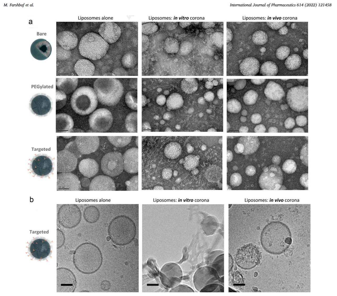
Picture 46. Above. Morphological and structural characterization of liposomes before and after protein corona formation in vitro and in vivo by electron microscopy. (a) Negative-staining TEM imaging showing the interaction of plasma proteins with naked, PEGylated, and targeted liposomes recovered from the bloodstream of CD-1 mice 10 minutes after injection and after in vitro incubation with isolated CD-1 mouse plasma; and (b) Cryo-EM imaging of targeted liposomes before and after their in vitro and in vivo interaction with plasma proteins. All scale bars are 50 nm.
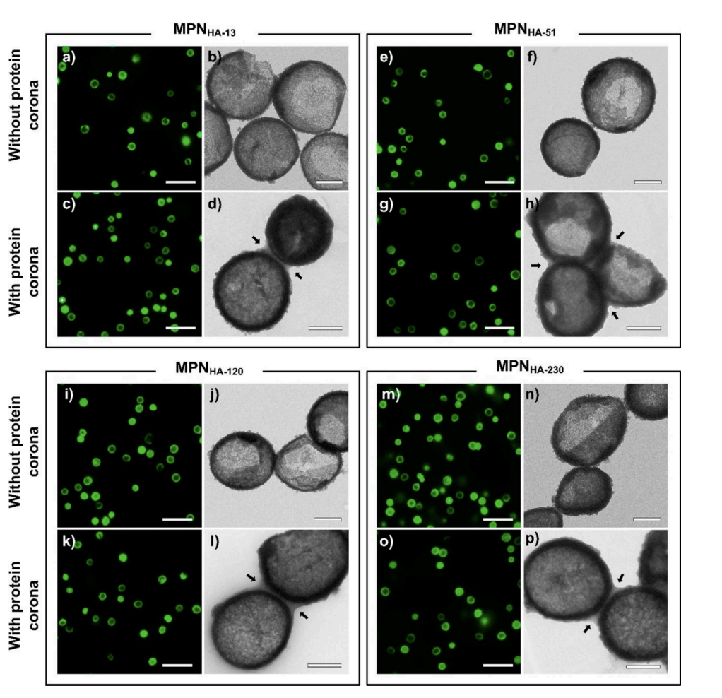
Picture 47. Above. Fluorescence microscopy images (a, c, e, g, i, k, m, o, scale bars are 5 μm) and TEM images (b, d, f, h, j, l, n, p, scale bars are 0. 5 μm) of MPNHA-13, MPNHA-51, MPNHA-120, and MPNHA-230 capsules without (top rows) or with (bottom rows) protein corona after incubation with human serum for 1 h. 13, 51, 120, and 230 represent the molecular weight of hyaluronic acid (HA). Arrows in TEM images (d, h, l, p) indicate protein corona around the capsules. Adapted with permission from ref (Ju et al., 2016). Copyright 2017 American Chemical Society.
“Nanoscale characterization of the biomolecular corona by cryo-electron microscopy, cryo-electron tomography, and image simulation” [6]
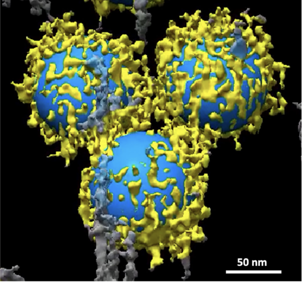
Picture 01. Above. TEM images of soft and hard BCs on the surface of NPs. Clusters of biomolecules are visible on both the carbon support film of the Cu TEM grid and the surface of the NPs before washing (a, b), and in the hard corona of NPs after washing three times (c, d). Higher-magnification images (b, d) of the surface of a single NP shows the clusters of biomolecules in the hard corona. Note the absence of larger clusters in the background of the unwashed sample.
Picture 03. Above. Representation of distribution pattern of clusters of biomolecules of the surface of individual NPs. The image is a snapshot generated from the 3D volume of Supplementary Movie 4.
Picture 04. Above. Selected slices from the reconstructed tomographic 3D volume (see Supplementary Figs. 3, 4, and Supplementary Movies 5–23 of the Supplementary Information; see the image analysis section for details of the generation of the 3D corona decoration). The 2D images represent virtual slices through the reconstructed 3D volume of the NPs and corona, from top to bottom. Local distribution and association of the biomolecules within the corona on the surface of the NPs are visible. The biomolecules appear as a dark dense structure bound to the surface of the NPs and at distances from the surface. Changes in the appearance of the individual dots from one image to the next image (see arrows) indicate that the size of these dots, representing biomolecules, is ~1 nm. The scale bar for all images in the panel are the same (100 nm).
“Graphene oxide–metal nanocomposites for cancer biomarker detection”. [12]
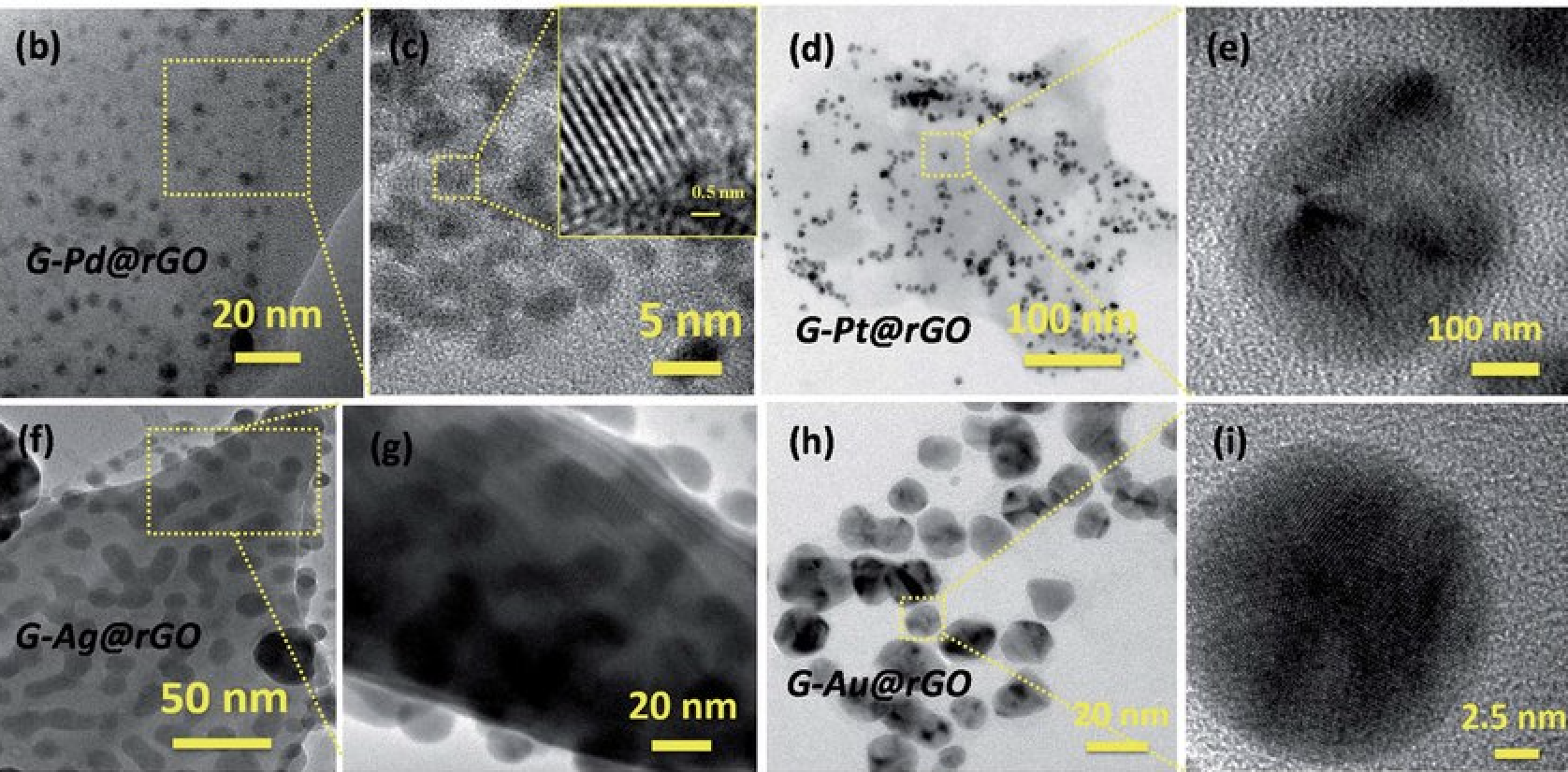
Picture 09. Above. (a) Formation of protein-corona on the surface of mNPs@rGO sheets. (b and c) G-Pd@rGO nanocomposite, inset of (c) shows a highresolution image of G-Pd@rGO, (d) G-Pt@rGO nanocomposite, (e) magnified image of G-Pt@rGO nanocomposite. (f and g) G-Ag@rGO nanocomposite and (h and i) G-Au@rGO nanocomposite.
“An environmental ecocorona influences the formation and evolution of the biological corona on the surface of single-walled carbon nanotubes”. [24]
According to their introduction. «Nanomaterials (NMs) taken up from the environment carry a complex ecocorona consisting of dissolved organic matter. An ecocorona is assumed to influence the interactions between NMs and endogenous biomolecules and consequently affects the formation of a biological corona (biocorona) and the biological fate of the NMs. This study shows that biomolecules in fish plasma attach immediately (within <5 min) to the surface of SWCNTs and the evolution of the biocorona is a size dependent phenomenon. Quantitative proteomics data revealed that the nanotube size also influences the plasma protein composition on the surface of SWCNTs. The presence of a preattached ecocorona on the surface of SWCNTs eliminated the influence of nanotube size on the formation and evolution of the biocorona. Over time, endogenous biomolecules from the plasma partially replaced the preattached ecocorona as measured using a fluorescently labelled ecocorona. The presence of an ecocorona offers a unique surface composition to each nanotube. This suggests that understanding the biological fate of NMs taken up from the environment by organisms to support the environmental risk assessment of NMs is a challenging task because each NM may have a unique surface composition in the body of an organism. ».
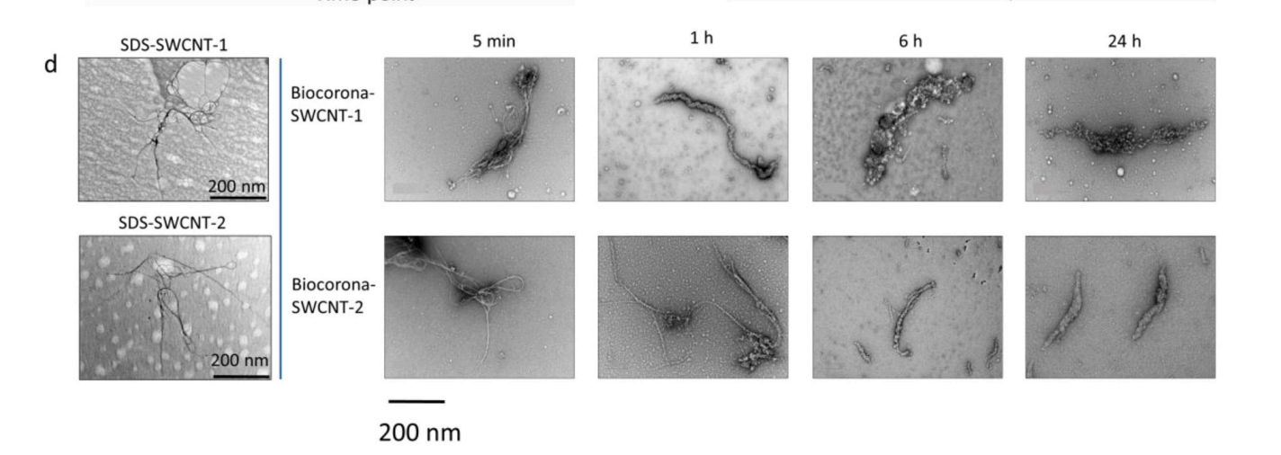
Picture 15. Above. a) Scheme of the incubation of SWCNTs in a suspension of dissolved organic matter (DOM). The brown molecules on the surface of the SWCNTs represent the DOM. b) Scheme of the incubation of SWCNTs with fish plasma. c) It shows the thickness of the biocorona-SWCNT complexes, which was measured (mean and SD of 100 tubes) using transmission electron microscopy (TEM). d) The TEM pictures were provided for dispersions of SWCNTs in 1% sodium dodecyl sulfate solution before incubation in plasma. For both SWCNTs, the biocorona increased the thickness with incubation time. e) Biological corona quantification by using protein as proxy. The figure shows the quantification of plasma proteins [femtogram (fg) per nanotube] at the indicated time points (values are mean ± SD).
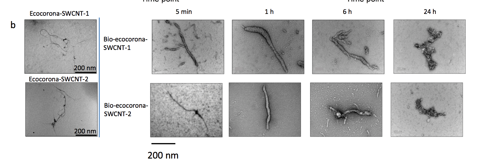
Picture 16. Above. Thickness of bio-ecocorona-SWCNT complexes, which was measured using TEM (mean ± SD, 100 complex) over time. b) TEM images showing the evolution and structure of the bio-ecocorona formed on the surface of the SWCNTs. c) Biocorona quantification by using the plasma proteins as proxies. The mass of the proteins on the surface of the SWCNTs (femtogram per nanotube) was measured at the indicated time points.
“The impact of nanoparticle protein corona on cytotoxicity, immunotoxicity and target drug delivery”. [8]
According to their introduction. «In a perfect sequence of events, nanoparticles (NPs) are injected into the bloodstream where they circulate until they reach the target tissue. The ligand on the NP surface recognizes its specific receptor expressed on the target tissue and the drug is released in a controlled manner. However, once injected in a physiological environment, NPs interact with biological components and are surrounded by a protein corona (PC). This can trigger an immune response and affect NP toxicity and targeting capabilities. In this review, we provide a survey of recent findings on the NP–PC interactions and discuss how the PC can be used to modulate both cytotoxicity and the immune response as well as to improve the efficacy of targeted delivery of nanocarriers». [8]
“Measurements of heterogeneity in proteomics analysis of the nanoparticle protein corona across core facilities” [14]
Picture 13. Above.Electron microscopy characterization. TEM images of (a–c) bare PSNPs, (d) protein corona-coated PSNPs that clearly reveal proteins on their surface after incubation with human plasma proteins, (e) and (f) Cryo-TEM images of protein corona-coated PSNPs obtained with a direct electron detector and phase plate, clearly showing the distribution of proteins on the surface of the PSNPs (black dots in the images (e, f) are 10 nm gold fiducial markers). The results are representative of three independent measurements.
“Platinum Nanoparticles As A Therapeutic Agent Against Dextran Sodium Sulfate-Induced Colitis In Mice”. [9]
Picture 08. Above. Transmission electron microscopy (TEM) images of platinum nanoparticles (PtNPs): (A) Pt-5nm; (B) Pt-30nm; (C) Pt-70nm.
“Formation of a protein corona influences the biological identity of nanomaterials”. [15]
« Biological dynamics can impact PC formation on NPs. One dynamic aspect is termed the “Vroman effect”, originally studied by Leo Vroman. The Vroman effect describes the phenomenon that certain proteins that initially associate with the nanoparticle PC, over time, within minutes or hours, are exchanged with a new set of proteins that possess higher affinities for the nanoparticle’s surface or the corona. This trafficking of proteins may occur within the SC in seconds to minutes due to the SC proteins’ low affinity for each other and for the HC proteins. In contrast, components of the HC are strongly bound to NPs and may take hours to exchange with higher affinity proteins, if at all ».


Voici une explication claire présentée par l’étude, de 2019, intitulée “Understanding the Chemical Nature of Nanoparticle – Protein Interactions”. [16]
«In the past decade, it has been rigorously established that all particles administered into biological fluids are inevitably and immediately (>0.5 min) coated by biomolecules, such as enzymes, proteins, peptides, and/or amino acids, to form what is known as a “corona” on the particle surface. The protein corona was first described in 2007 by Dawson et al., who studied the proteins associated with nanoparticles (NPs) with a wide range of binding affinities in various biological fluids. The protein corona evolved to the “biomolecular corona” that surrounds NPs in 2012 by Monopoli et al.
The formation process of the biomolecular corona is dynamic and depends on the unique properties of the NPs, such as size, surface chemistry, ligands, nature, shape, charge, surface curvature, and environmental impact. The nature and composition of the biomolecular corona around NPs may affect the biological fate, cellular uptake, immunological response, and biodistribution of NPs. After administration into the bloodstream, the corona composition of NPs dynamically shifts until an equilibrium reached. Dramatic changes take place on a “soft corona” due to the low affinity of proteins, while only small changes occur in a “hard corona”, where comparably stronger interactions bind the biomolecules onto a NP’s surface. The hard corona achieves equilibrium in seconds to 30 min, while a soft corona may require several hours. Nowadays, most analytical techniques focus on studying the hard corona; only a few examine the soft corona with real-time monitoring.
Understanding the unique properties of the protein corona is essential to nanomedicine. This protein coating may slow the degradation of NPs, decrease their cytotoxicity, influence their in vivo half-life, change their uptake, stimulate their internalization by cells, and affect the inflammation signaling pathway. Due to the complexity of biomolecular coronas, it is essential that one uses appropriate and accurate methods to characterize the corona so as to understand the adsorption process, adsorption kinetics, and binding affinity, as well as quantify the adsorbed biomolecules. In addition, the nanoparticle−biology (nanobio) interface characterization is an important consideration. Human plasma consists of more than 3500 proteins, but not all of them will adsorb onto the surface of a given NP. The protein coronas formed around NPs possess different natures and usually endow the NPs with new biological effect and identity».

“A comprehensive assessment of microbiome diversity in Tenebrio molitor fed with polystyrene waste”. [4]
Toxicity of nanoplastics. «Nanoplastics cause toxic effects similar to those induced by microplastics. Reproductive disorders, alteration of the immune system and developmental changes have been described in crustaceans, echinoderms and mollusks exposed to nanoplastics. However, the toxic mode of action of nanoplastics may be related to their behavior in biological fluids, which is comparable to that of engineered nanoparticles (defined as “intentionally produced for commercial purposes to have specific properties or a specific composition”. It has been well described that nanoparticles spontaneously adsorb biomolecules from biological fluids, forming a dynamic molecular layer called a “protein corona”.
The protein corona modulates the fate and toxicity of nanoparticles in organisms via interaction with cellular uptake mechanisms, the innate immune system and detoxification processes. Although research on nanometric materials and proteins has mainly focused on metal (silver) and metal oxide (zinc oxide, titanium dioxide) nanoparticles, polystyrene nanoparticles also induce the formation of a protein crown».
I can only underline the importance of what the author evokes with regard to what I have called “the necromolecular corona” of graphene… or of any other metallic nano-particle.
Whether it is graphene, silver, cobalt, titanium, boron, etc. nanoparticles, the necro-molecular corona is, resolutely, a corona of spikes, spicules, points, razor blades that shred blood cells, neuronal cells… and that graft themselves to them – when they survive.
Other Pictures of Necro-Molecular Coronas
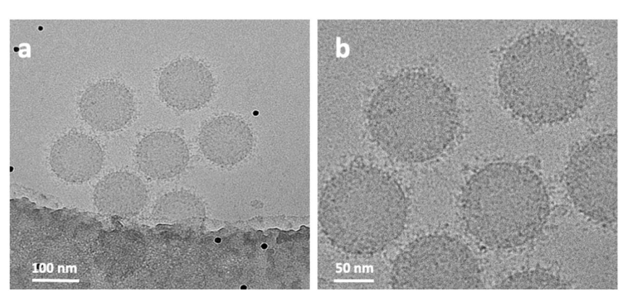
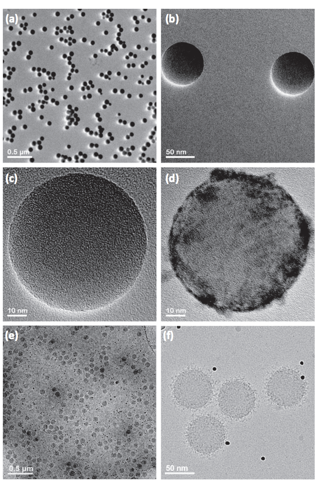
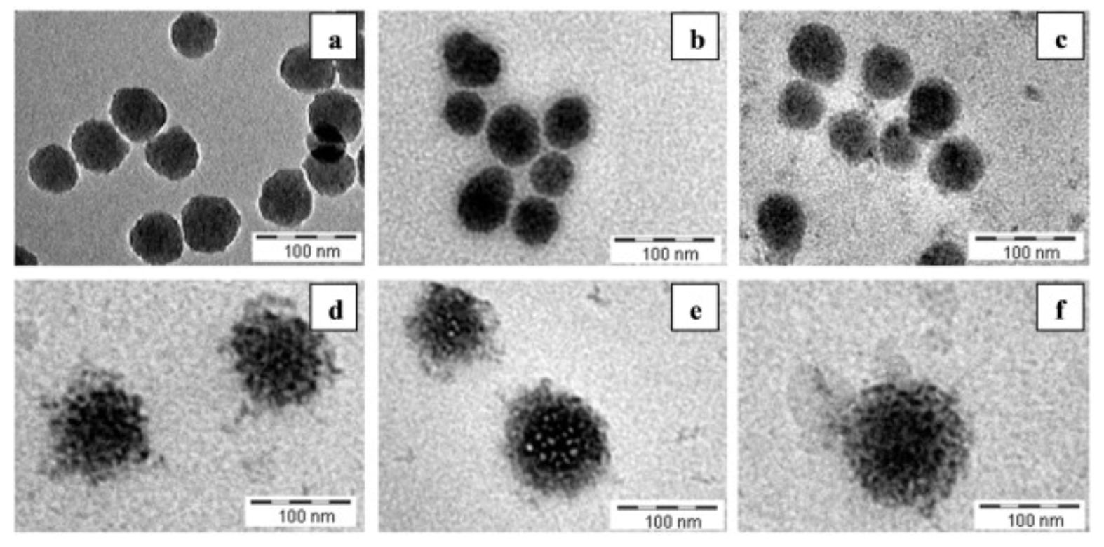
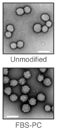
Picture 10. [10] Sars-CoV-2 and graphene oxide necro-molecular coronas.

Picture 11. v Fullerenes. [13]
Picture 12. Above. Transmission electron microscopy image showing the formation of a biomolecular crown around the surface of the nanoparticles. Credit: Morteza Mahmoudi, Brigham and Women’s Hospital.
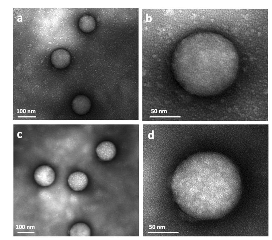
Picture 14. Above. Stability of silica nano-particles as a function of time and formation of a protein corona in a biological medium. Concentration of 1×10¹² nanoparticles mL-1, incubation times: (a) 0 min, (b) 10 min, (c) 6 h, (d) 48 h, (e) 1 week, (f) 2 weeks in DMEM + 10% FBS.
Picture 17. Above. Carbon nanotubes and protein coronas in human blood. [25]
Picture 18. Above.The lower panel shows a cluster of short-cut SWCNTs (single arrow) entrapped in chromatin fibers (two arrows) of purified neutrophil extracellular traps [see Farrera et al6 for further details]. SEM courtesy of K. Hultenby, Karolinska Institutet. 26]
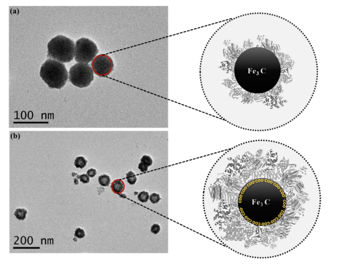
Picture 19. Above. TEM images of (a) non-coated Fe2C NPs and (b) COOH coated Fe2C NPs, while zoomed images show possible protein corona. [27]
Picture 20. Above. The lower panel shows a cluster of short-cut SWCNTs (single arrow) entrapped in chromatin fibers (two arrows) of purified neutrophil extracellular traps [see Farrera et al6 for further details]. SEM courtesy of K. Hultenby, Karolinska Institutet.. [17]
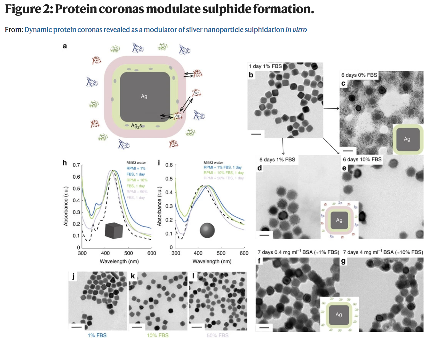
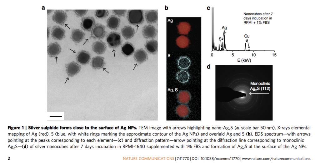
Picture 39. Above. Molecular coronas modulate the sulfidation of silver nanoparticles in vitro.
Alleged Pictures of the invisible Sars-CoV-2
These are only a few photographs, which one finds on the Web representing, supposedly, the Sars-CoV-2. They are very blurred… and could, really, represent many things.
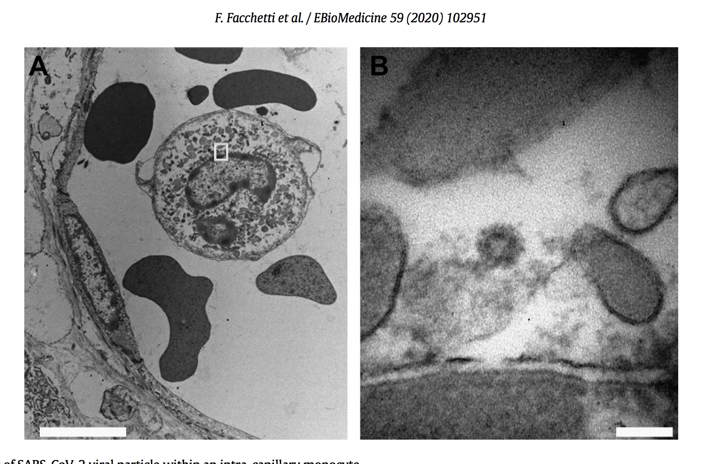
Picture 30. Electron microscopy of SARS-CoV-2 viral particle within an intra-capillary monocyte. (A, B) In the cytoplasm of an intra-capillary monocyte (A, bar 5 mm) a particle morphologically consistent with coronavirus is present (B, high magnification of the area shown in A, bar 100 nm). [18]
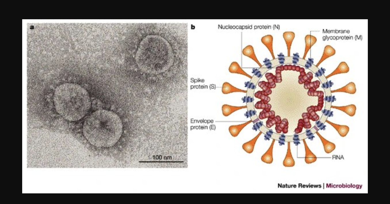
Photo 31. [19]
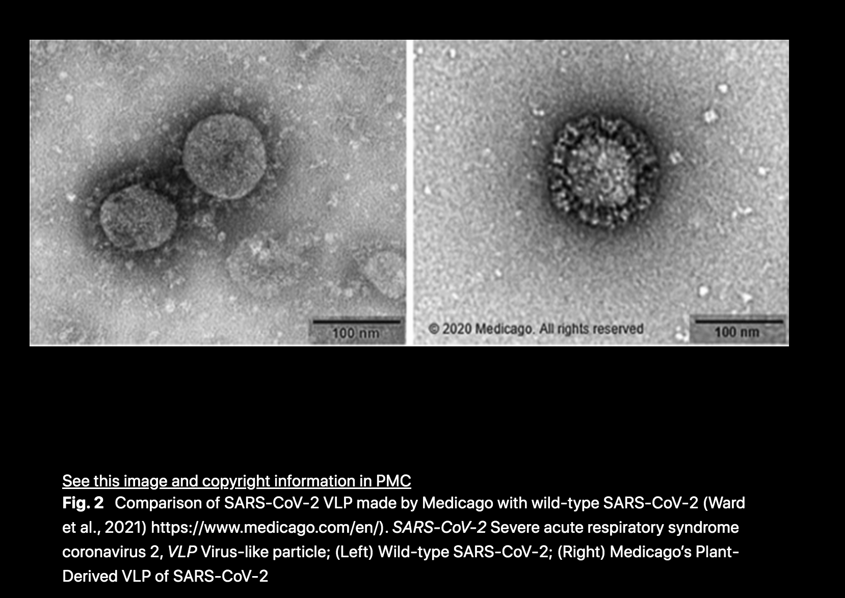
Photo 35.
Picture 37. Transmission electron microscopy analysis of SARS-CoV-2 infected intestinal organoids: (a to h) overview of an intact organoid (a) showing the onset of virus infection (b to d) at different stages of the viral lifecycle, i.e., early double membrane vesicles (DMVs); (e), asterisk, initial viral production in the Golgi apparatus (f and g) and complete occupation of virus particles inside the endomembrane system (h); (i to k) extracellular viruses are observed in the lumen of the organoid (i), and are found at the basal (j) and apical side (k) alongside the microvilli (arrows). Scale bars – 10 μm (a), 2.5 μm (b to d), 250 nm (e), (f), and (h to k) and 100 nm (g). (l to q) Overview of an organoid (l) showing severely infected cells (m and o), disintegrated cells (o) and stressed cells as evident from the atypical nucleoli (p); intact cells reveal DMV areas of viral replication (p), asterisks, and infected Golgi apparatus (q); (r) extracellular clusters of viruses. Scale bars – 10 μm (l), 2.5 μm (m to p) and 250 nm (p to r). Image credit: Lamers et al, doi: 10.1126/science.abc1669.

Picture 38. Above. TEM image of SARS-COV-2 infected Vero cells from the Electron Microscopy Unit, Research Technologies Branch, Rocky Mountain Labs, NIAID.
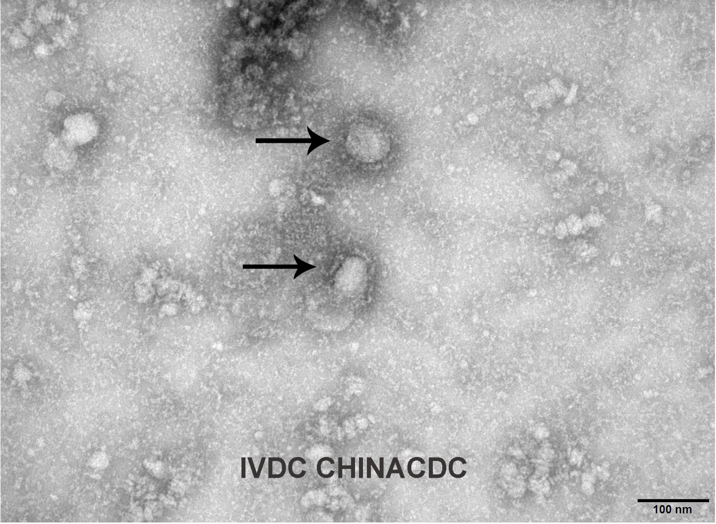
Picture 41. Above. A transmission electron microscopy image of the first isolated case of coronavirus, as obtained by Reuters on January 27, 2020. Courtesy of IVDC, China CDC via GISAID/Handout via Reuters.
It should be noted that George Gao, the director of the Chinese CDC, once stated – in an interview I reported at the time – that the Chinese had never isolated Sars-CoV-2.
Picture 48. Above. [36] Wuhan strain in negative stain (a) and cryo-ET (b). Alpha strain (B.1.1.7) in negative stain (c) and cryo-ET (d). Beta strain (B.1.351) in negative stain (e) and cryo-ET (f). Delta strain (B.1.617) in negative stain (g) and cryo-ET (h). The negative stain fields show predominantly circular views of virions, while the tomogram sections show circular, elliptical side, and oblique views. All four variants show similar virion profiles decorated with Spike proteins. We have measured the three principal axis dimensions. The cylinder cross section is slightly elliptical, and the cylinder height (Wuhan average = 39 nm) is less than half that of the average diameter (Wuhan average = 102 nm). Similar shapes and dimensions were observed and measured from cryotomograms for the Alpha, Beta and Delta variants.
According to this study, the proposed diameter of the CoqueVide strain made in Wuhan is 102 nm – three times less than the Delta-Omicron variant, of Professor Didier Raoult in France, presented in the beginning of this essay.
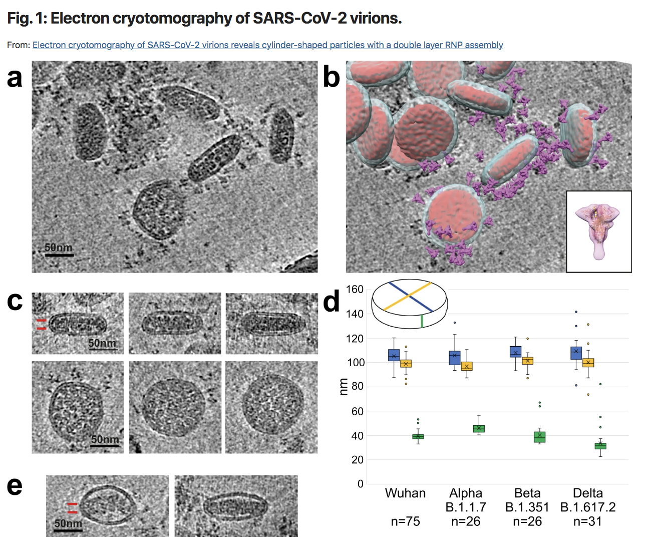
Picture 49. Above. [36] a Tomogram section of Wuhan virions in cross-section showing surface spike proteins surrounding the viral membrane bilayer and the nucleocapsid in the virus interior. b Same field of view with models for the virus membrane (pale blue), nucleocapsid (red), and the spike glycoprotein (purple) overlaid. The glycoprotein map (inset) was obtained by subtomogram averaging of spikes and fitted with a pre-fusion S model (pdbid 6zge). c Gallery shows cross-sections through narrow side views and circular top views of Wuhan virions. The side view shows two layers of density for RNP subunits in the nucleocapsid assembly (indicated by two red lines), which is separated from the top and bottom membrane bilayer. The circular top views show dense RNP subunits that extend to the bilayer. d Virion dimensions for 4 variants (Wuhan n = 75, Alpha n = 26, Beta n = 26, Delta n = 31). Box and whisker plot of the measurements of 3 orthogonal dimensions as shown in inset: circular view major axis (blue), circular view minor axis (yellow), side view (green). The boxes represent the interquartile range of measurements, line across box = median, x inside box = mean (see Supplementary Table 1 for values), whiskers extend to minimum and maximum measurements, circles = outliers. e Wuhan virions of aberrant ellipsoidal morphology show a two layer nucleocapsid, which abuts the membrane at the highly-curved sides while remaining separated from the membrane bilayers above and below.
Molecular Necro-Coronas in Humans and other Animals
“Unveiling the Bio‑corona Fingerprinting of Potential Anticancer Carbon Nanotubes Coupled with d‑Amino Acid Oxidase”. [2]
It seems interesting to propose the entire abstract on the use of multiwall carbon nanotubes as nanovectors for the transfer of allegedly therapeutic substances, allegedly bio-compatible, generating a necro-crown, biologically-metallic. CoV-2.
« The oxidation therapy, based on the controlled production of Reactive Oxygen Species directly into the tumor site, was introduced as alternative antitumor approach. For this purpose, d-amino acid oxidase (DAAO) from the yeast Rhodotorula gracilis, an enzyme able to efciently catalyze the production of hydrogen peroxide from d-amino acids, was adsorbed onto multi-walled carbon nanotubes (MWCNTs), previously functionalized with polylactic-co-glycolic acid (PLGA) or polyethylene glycol (PEG) at diferent degrees to reduce their toxicity, to be targeted directly into the tumor. In vitro activity and cytotoxicity assays demonstrated that DAAO-functionalized nanotubes (f-MWCNTs) produced H2O2 and induced toxic efects to selected tumor cell lines. After incubation in human plasma, the protein corona was investigated by SDS-PAGE and mass spectrometry analysis. The enzyme nanocarriers generally seemed to favor their biocompatibility, promoting the interaction with dysopsonins. Despite this, PLGA or high degree of PEGylation promoted the adsorption of immunoglobulins with a possible activation of immune response and this effect was probably due to PLGA hydrophobicity and dimensions and to the production of specifc antibodies against PEG. In conclusion, the PEGylated MWCNTs at low degree seemed the most biocompatible nanocarrier for adsorbed DAAO, preserving its anticancer activity and forming a bio-corona able to reduce both defensive responses and blood clearance.». Emphasis from Xochi.
Bottom line. This summary is full of: generally, seems, possible, probably… It is fake science because with graphene, it is better to pretend that all is well in the best of all worlds of bio-compatibility.
Why? Because, quite simply, if carbon nano-tubes are able to destroy cancer cells it is because they are toxic, intrinsically. Therefore, the demented scientists have to bypass the barriers and generate a necro-corona, biologico-metallic, able to break the resistances in order to go to the heart of the tumor… while claiming bio-compatibility.
This study highlights that these chimeric necro-coils – generated by multi-walled carbon nanotubes – contain retinoid linkages, heparin linkages, arylesterases, RNA linkages, protease linkages, hemoglobin linkages, serine proteases, endopeptide inhibitors, serpins, calcium ion binding, peptidase inhibitors, retinol binding proteins, homodimerization proteins, metal ion binding, copper ion binding, hydrolases, ferroxidases, thyroid hormone binding, scavenger receptors, apolipoproteins…
“Toxicity mitigation and biodistribution of albumin corona coated graphene oxide and carbon nanotubes in Caenorhabditis elegans”. [4]
« In this work, the toxicity and biodistribution of graphene oxide (GO) and oxidized multi-walled carbon nanotubes (MWCNT) were investigated in Caenorhabditis elegans. Bovine serum albumin (BSA) was selected as a model protein to evaluate the influence of protein corona formation on materials physicochemical properties, colloidal stability, and toxicity. Biological assays were performed to assess the effects of bare and albumin corona coated materials on survival, oxidative stress, intestinal barrier permeability, growth, reproduction, and fertility. Critical alterations in topography, surface roughness and chemistry of GO and MWCNT were observed due to albumin corona formation. These modifications were associated with changes in colloidal stability of materials and prevention of their aggregation and sedimentation in nematode testing medium. Both GO and MWCNT caused damage to nematode survival, growth, reproduction, and fertility, as well as enhanced oxidative stress and permeability of the intestinal barrier. But GO was more toxic than MWCNT to C. elegans, especially at long-term assays. Albumin corona mitigated 100% of acute and chronic effects of MWCNT. In contrast, the negative effects of GO were not completely mitigated; GO inhibited 16.2% of nematode growth, 86.5% of reproduction, and 32.0% of fertility at the highest concentration evaluated (10 mg L-1), while corona coated GO mitigated 50% and 100% of fertility and growth, respectively.
Confocal Raman spectroscopy imaging was crucial to point out that bare and albumin corona coated GO and MWCNT crossed the C. elegans intestinal barrier reaching its reproductive organs. However, BSA corona protected the nematodes targeted organs from negative effects from MWCNT and blocked its translocation to other tissues, while coated GO was translocated inside the nematode affecting the functionality of crucial organs. In addition, coated MWCNT was excreted after 2 h of food resumption, whereas coated GO still accumulated in the nematode intestine. Our results demonstrate that the materials different translocation and excretion patterns in C. elegans had a relation to the impaired physiological functions of primary and secondary organs. This work is a contribution towards a better understanding of the impacts of protein corona on the toxicity of graphene oxide and carbon nanotubes; essential information for biological applications and nanosafety.»
In conclusion, the toxicity of oxidized multiwall carbon nanotubes and graphene oxide differed, but both caused damage to nematode survival, growth, reproduction, and fertility, as well as increased oxidative stress and gut barrier permeability.
Molecular Necro-Coronas in Plants
“Bio- and eco-corona related to plants: Understanding the formation and biological effects of plant protein coatings on nano-particles”. [3]
The ever-growing agriculture based on nanotechnologies is an agriculture that integrates metallic nanoparticles, in particular Graphene derivatives, into the synthetic fertilizers and biocides of conventional agriculture.
«The thriving nano-enabled agriculture facilitates the interaction of nanomaterials with plants. Recently, these interactions and their biological effects are receiving increasing attention. Upon entering plants via leaves, roots, stems, and other organs, nanoparticles adsorb numerous biomolecules inside plants and form bio-corona. In addition, nanoparticles that enter plants through roots may have formed eco-corona with root exudates in the rhizosphere environment before contacting with plant exogenous proteins. The most significant biological effects of plant protein corona include changes in protein structure and function, as well as changes in nanoparticle toxicity and targeting ability. However, the mechanisms, particularly how protein corona affects plant protein function, plant development and growth, and rhizosphere environment properties, require further investigation. Our review summarizes the current understanding of the formation and biological effects of nanoparticle-plant protein corona and provides an outlook on future research.». Les soulignements sont de mon fait.
In conclusion, necro-crowns are also formed in plant tissues and in the rhizosphere. Metallic nanoparticles are toxic for plants and their environment.
It is worth noting the unfortunate, and misleading, expression “eco-crowns” – which are formed by the fusion between metallic nano-particles and root exudates.
Molecular Necro-Coronas in Food
“Investigation of the interactions between food plant carbohydrates and titanium dioxide nanoparticles”. [5]
This study focuses on titanium dioxide nanoparticles that are commonly used as a food whitening agent in candies, chocolates and high-carbohydrate cakes.
« Titanium dioxide (TiO2) is commonly used as food whitening in candies, chocolates, and cakes with high carbohydrate contents. The potential interaction between the food carbohydrate and food grade TiO2 nanoparticle was little known. Therefore, we explored the interaction between TiO2 nanoparticles and seven common carbohydrates, including monosaccharides, disaccharides, and polysaccharides. The result showed that all the carbohydrates tested interacted with the surfaces of the TiO2 nanoparticles and formed biocoronas. TEM and SEM images provided information about the morphology formation of biocoronas. The surface potential and size of the TiO2 nanoparticles altered after interacting with the carbohydrates. FTIR spectroscopy and QCM-D findings showed insights into the molecular origin and nature interaction between TiO2 and carbohydrates. The results illustrated that TiO2 nanoparticles can interact with carbohydrates, enter the body as a food additive, and interact with food matrix for a series of reactions. Compared with monosaccharides or disaccharides, food polysaccharides have stronger adsorption on the surface of nanoparticles. This is a preliminary judgment for the subsequent in vitro simulated digestion. Our result could be useful for understanding and controlling the behavior of nanoparticles in food and the human gut. ».
Bottom line. The results showed that nanoparticles titanium dioxide can interact with carbohydrates, enter the body as a food additive and interact with the food matrix for a series of reactions.

![[10]](https://nobulart.com/wordpress/wp-content/uploads/2021/07/sarsgo.jpg)